
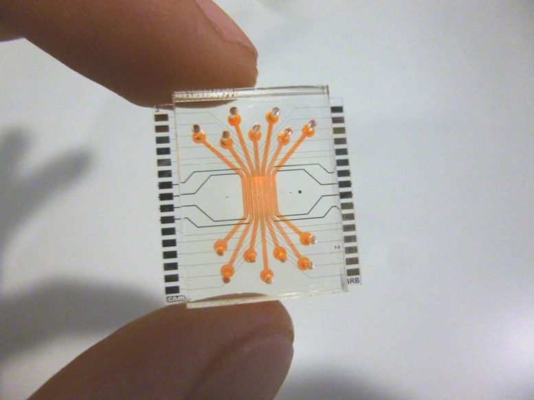
The new chip developed by Barcelona researchers. Credit: Jose Yuste (CSIC-IMB-CNM)
For some years, scientists have been researching in order to find ways of reducing animal testing and accelerating clinical trials. In vitro assays with living cells is an alternative, but it has limitations as the interconnection and interaction between cells cannot be easily reproduced.
To overcome the latest, scientists are developing systems that mimic tissues and organ functions in conditions very close to reality. These type of devices, called “Organ-on-a-chip”, include microenvironments and microarchitectures in order to emulate the living organs and tissues.
A team of scientists in Barcelona has developed a microfluidic device that mimics the human blood-retinal barrier. The scientists are from the Biomedical Applications Group of the Microelectronics Institute of Barcelona (IMB-CNM) of the Spanisch Council for Scientific Research (CSIC), the CIBER-BBN, and from the Diabetes and Metabolism group of the Vall d’Hebron Research Institute (VHIR), CIBERDEM- Institute of Health Carlos III, and the Universitat Autònoma de Barcelona (UAB).
The study, featured on the cover of the journal Lab-on-a chip, is what experts technically call “proof of concept”, that is, it demonstrates the feasibility of the design imagined by the scientists.
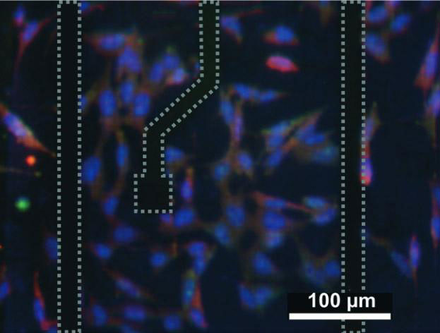
Cells on the chip. Credit: José Yeste. (CSIC-IMB_CNM)
Emulating the structure of the blood-retinal barrier
José Yeste, a CSIC scientist at the IMB-CNM and at the Microelectronics and Electronics Systems Department at the UAB, and main author of the study, explains that the device is composed by several parallel compartments, arranged to emulate the retinal layer structure. In every compartment a type of cells has been cultured: endothelial cells (which constitute capillary vessels which carry oxygen and nutrients), neuronal cells (which form the neuroretina), and retinal pigmented epithelial cells, which form the outer layer of the blood-retinal barrier.
The compartments are interconnected by a grid of microgrooves under the cells, with which cells can exchange signal molecules and therefore communicate between them. As a result, substances produced by some cells can reach the other cells, generating a cellular communication and interaction like in a living organ. Also, the device enables to expose endothelial cells to the particular mechanical conditions, like the ones induced by the blood stream.
As Rosa Villa, a CSIC scientist and leader of the Biomedical Applications Group, explains, “in the living organism, endothelial cells that cover the inner walls of blood vessels are exposed to the mechanical stimulus of the blood stream. In the cell cultures where this condition is not reproduced, we could say the cells are like “sleepy” and do not react as they would do in real conditions”.
Rafael Simó, who leads the Diabetes and Metabolism group of the Vall d’Hebron Research Institute (VHIR) says: “The most relevant characteristic of this technology is that mimics what happens ‘in vivo’ in the retina and therefore can be an essential tool to boost the in vitro experimentation. On the device, the cells grow constantly in contact with a fluid, as it happens in the human retina. Also, the cells keep a close interaction between them by chemical mediators, which makes possible to see what happens in a type of cell when another type of cell nearby is harmed. Also, it is possible to measure electrical resistance for assessing the functionality of the retinal neurons”.
The scientists tested the correct formation of the blood-retinal barrier assessing its permeability, its electrical resistance and the expression of some proteins of the tight junctions between cells, which are expressed when cells have established a barrier function.
The tests have been designed to check whether the barrier was properly formed, but keeping the natural permeability to allow the pass of nutrients and oxygen, and to find out if the cells were in contact and interacting.
This device, scientists say, can be used to study the effects of molecules or harming conditions on the human retina. The team also wants to use the device to study the diabetic retinopathy, a disease whose causes and progression are not well understood yet.
Previously, the team lead by Rosa Villa at the Microelectronics Institute of Barcelona (IMB-CNM) of the CSIC did create a device that emulates the blood-brain barrier.
Also, they developed a microfluidic chamber (Liver-on-a chip) that mimics the hepatic microcirculation. In this case, they designed and manufactured the device together with scientists at the IDIBAPS.


